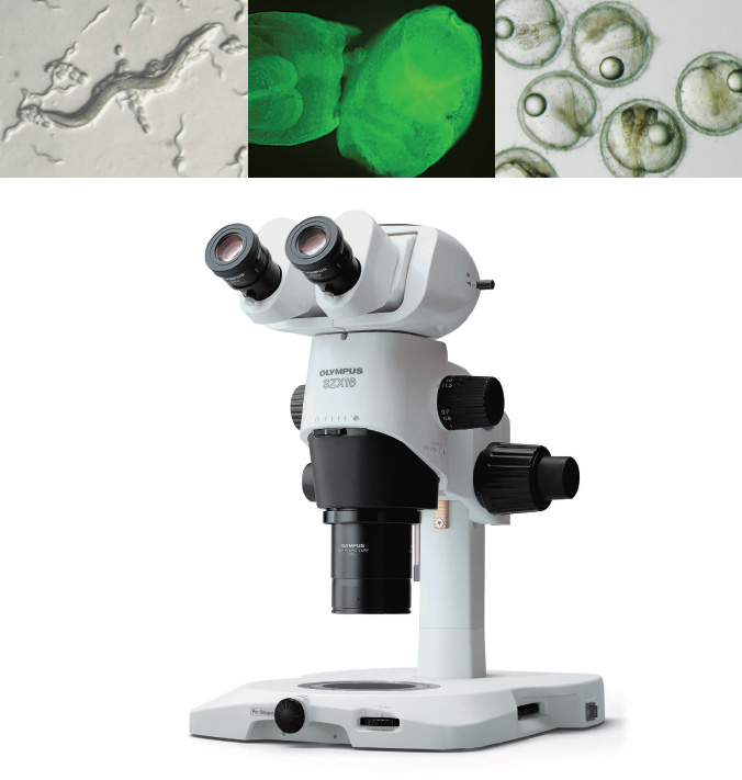
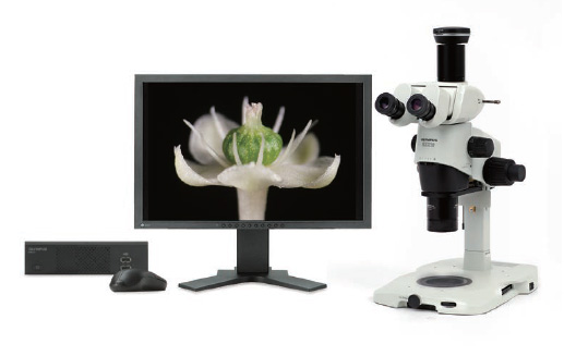
Olympus SZX2 series stereo microscopes are up to the challenge of leading-edge microscopy applications, offering an exceptionally wide zoom ratio and high numerical aperture (NA). Excellent image clarity and a flexible optical system make the SZX2 series easy to use, while their advanced optics, improved functionality, and ergonomic design deliver an outstanding user experience. Modern life science laboratories require the most effective imaging tools to observe a vast quantity of live specimens. The SZX2 stereo microscope series is designed to meet these needs and is refined to the highest levels of quality and performance. The combination of a high NA and a multi-wavelength, astigmatism-free design yields high-resolution images with an increased depth of field. Furthermore, the quad-position LED transmitted light illumination base enables you to easily switch the observation method and contrast level by changing cartridges. The SZX2 microscope is redesigned with improved ergonomics that reduce operator fatigue and enable comfortable observation over a long period of time.
Sharp Images That Enhance Your Research
Setting the Standard in Image Clarity
The microscope’s multi-wavelength, astigmatism-free design effectively eliminates image-deforming aberrations, enabling remarkably sharp 3D imaging and enhanced specimen manipulation. With an apochromatic lens system that effectively reduces chromatic aberration, the latest proprietary SZX16 optical system provides vivid 3D observation images of various specimens.
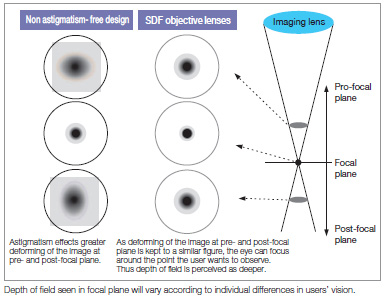
Sharp, Detailed Observation of Specimens
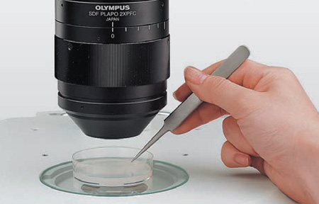
By reducing astigmatism, SDF objective lenses prevent the image deforming in the pre- and post-focal planes, offering a deeper depth of field. These design features enable stress-free use of forceps in the field of view during live sample selection and acquisition. When these objectives are combined with the transmitted light illumination base, users can observe low-contrast, transparent specimens. This reduces oversights for specimen selection, dissection, and manipulation.
Integrated Apochromatic System
The apochromatic system—integrated into the observation tubes, zoom body, and objectives—eliminates chromatic aberration throughout the zoom range and helps acquire high image quality without chromatic blur.
Optical Performance with Less Fatigue
A 360º view of balanced images is made possible by accommodating vertical and horizontal parameters. Discomfort in the eyes and body, as well as stress from long periods of observation or operation, are effectively reduced.
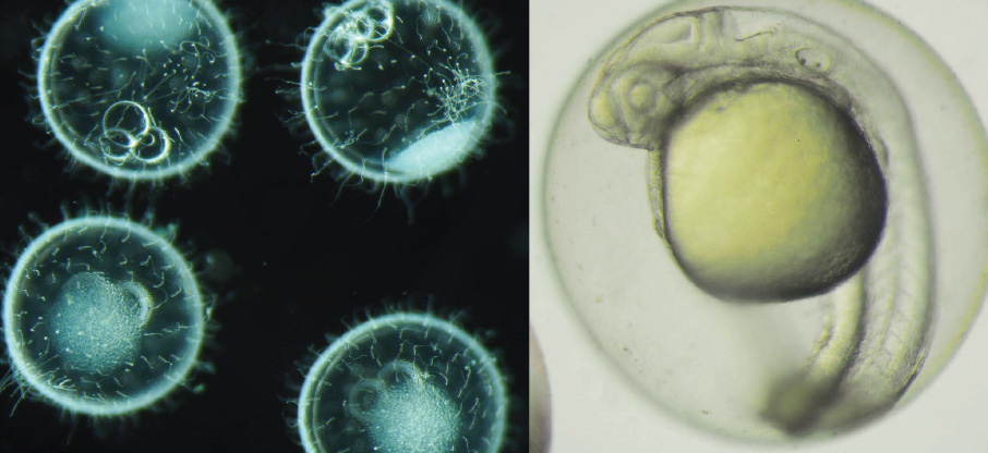
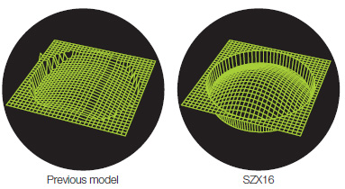
SZX16: Optics Easily Accommodate Thick Specimens
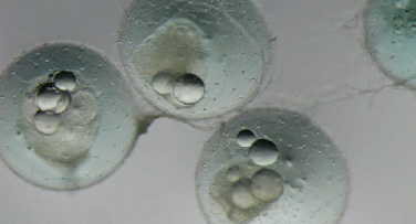
The ability to clearly perceive the depth and dimensions of thick specimens, such as eggs and embryos, is important in many applications. The SZX16 delivers clear 3D images from the surface and interior of live specimens for applications such as dissection.
Efficient Observation from Low to High Magnification,
Even in Fluorescence Imaging
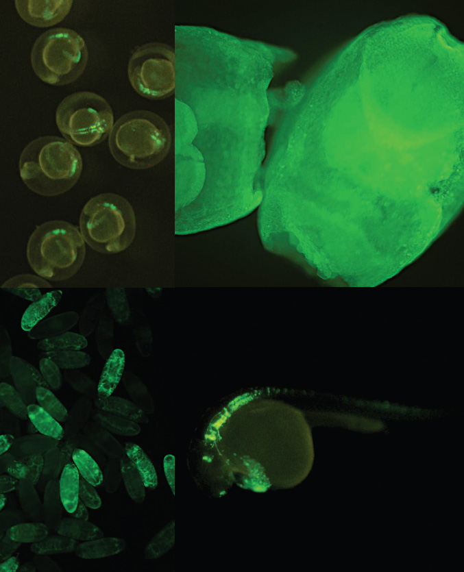
SDF Objectives Significantly Improve Signal Intensity and Support Bright Fluorescence Observation
Bright fluorescence observation is important in biological and medical research. Weak fluorescence is a common problem when observing specimens at low magnification under a stereo microscope. The SZX16 microscope enables even and bright fluorescence observation from low to high magnifications.
High NA for Bright Fluorescence Observation
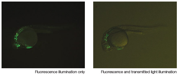
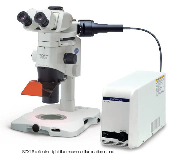
The SDF lenses’ high NA greatly improves fluorescence sensitivity. Furthermore, the newly designed near-vertical reflected light illuminator’s excitation light paths are independent from the observation paths, enabling substantially improved excitation light efficiency. These features provide far brighter fluorescence observation than conventional stereo microscopes at all magnifications. Transmitted light observation to verify the specimen’s outline is possible even under reflected light fluorescence observation.
Even and Seamless Fluorescence Observation
from Low to High Magnification
The near-vertical reflected light illuminator works in conjunction with the zoom function to provide even illumination over the entire magnification range.
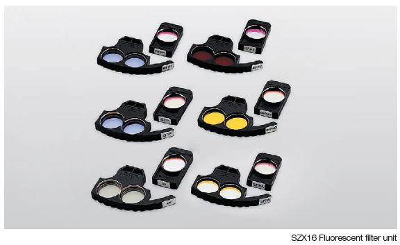
Five-Position Turret with Nine-Filter Selection
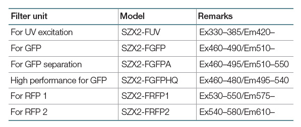
Six filter units, ranging from UV excitation to red fluorescent protein (RFP), enable imaging using various fluorescent dyes and proteins. Olympus high-quality (HQ) filters have an edge steepness and high transmission that efficiently detect the fluorescent light to enhance and capture brighter fluorescent images in precise detail.
Configure the Microscope to Match Your Needs
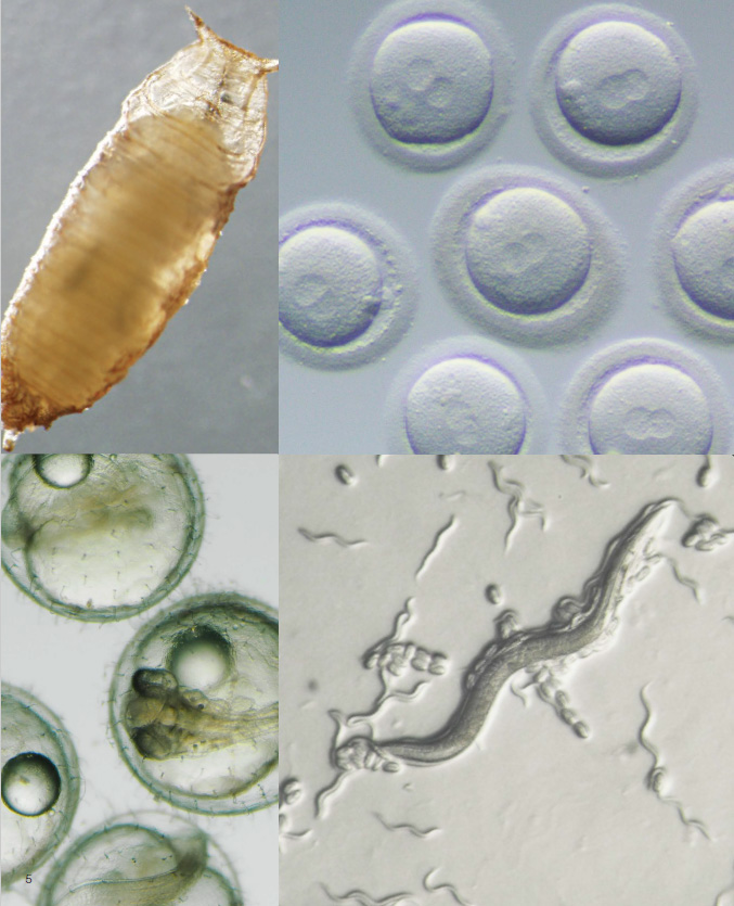
The SZX2 microscope handles a variety of specimens and operations—from large specimens such as mice to small ones, including Zebrafish, nematodes, C. elegans, or drosophila eggs—with an effective combination of high numerical aperture and wide working space. Moreover, the transmitted light illumination base is thin (only 41.5 mm (1.6 in.)) to provide a wide working space and enable multiple users to work comfortably.
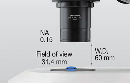
Wide Working Space and High NA
W.D. 60 mm and NA 0.15 from the 1X objective
The 1X objective has a 60 mm working distance that gives the user room to move and an NA of 0.15 that meets the needs of advanced research. Also available are 0.8X objectives that have a working distance of 81 mm, and provide not only a larger working space between objective lenses and sample but also a total magnification of 5.6x–92x (using the WHN10X-H).
Easy-to-access 2X objectives and correction collar
The intelligent design enables users to easily access objectives and delivers a high NA of 0.3 for easy specimen selection. An additional correction collar can adjust image quality independently of the specimen.
Ergonomically Designed, User-Friendly Base
Offering a wide working space in which users can place several Petri dishes, these illumination bases have an ergonomic, beveled design so users can work comfortably and naturally.
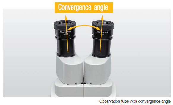
Observation Tube with Optimized Convergence Angle
Relieves Eyestrain
Working with an ophthalmologist, Olympus investigated and confirmed a correlation between stereo microscope optical systems and eyestrain. Specifically, the angle between right and left lines of vision (convergence angle) directly impacts eyestrain. The SZX2 series has an optimized convergence angle designed to enable users to make observations from a natural position that minimizes eye fatigue. This solution effectively eliminates eyestrain during long periods of observation.
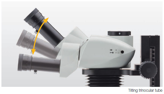
Ergonomic Accessories Enable Users to Optimize the
Microscope for Their Comfort
To improve the ergonomics of our stereo microscopes, Olympus introduced a long tilting trinocular tube (SZX2-LTTR). This trinocular can be adjusted from 5 to 45 degrees. In addition, the eyepoint adjuster (SZX2-EEPA) can raise and lower the eyepoint within a 120 mm range. Combining these units enables users to reduce stress and fatigue over longer periods of time by working in a natural posture. To improve the ergonomics of our stereo microscopes, Olympus introduced a long tilting trinocular tube (SZX2-LTTR). This trinocular can be adjusted from 5 to 45 degrees. In addition, the eyepoint adjuster (SZX2-EEPA) can raise and lower the eyepoint within a 120 mm range. Combining these units enables users to reduce stress and fatigue over longer periods of time by working in a natural posture.
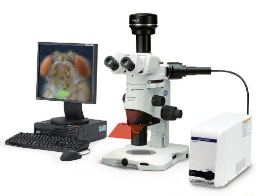
Reproduce True-to-Life Images with an Olympus Digital Camera
Each microscope digital camera in the SZX2 lineup captures images at high resolution. Olympus stereo microscopes and digital cameras contribute to leading-edge research in biology and medicine.
High-Performance Digital Cameras Provide Accurate and Detailed Image Capture (DP74/DP23)
DP74 Digital Camera
The DP74 color fluorescence camera captures realistic, high-quality images and has features that enable users to make their observations easily. With a wide field of view, operators can capture images of more of their sample, quickly. In applications like histology, the DP74 camera accurately reproduces colors to render natural images of your specimen. The camera displays a realistic image, so what appears on the monitor looks the same as what you see looking through the microscope’s eyepieces. Users can remain comfortable during their work since they can just watch the monitor rather than having to go back and forth between the monitor and eyepieces. The camera is easy to use, so it integrates into any workflow, making it simple to capture publication-quality images. *DP74 is not for clinical diagnostic use.
DP23 Digital Camera
The DP23 stand-alone camera smoothly displays live images in high definition while enabling easy observation, focusing, framing, and image archiving. Fine structures are precisely reproduced, and subtle color differences enable users to accurately identify targets on the monitor rather than having to look through the eyepieces. The dedicated control box provides smooth and intuitive operation using a touch screen monitor or a mouse (no PC required). *DP23 is not for clinical diagnostic use.
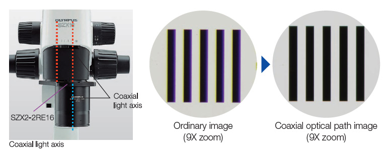
Vertical Observation
The revolving nosepiece matches the objective lens center to the zoom lens optical path for images with reduced aberration. Image shifting from focus change is eliminated for effective 3D rendering by software.
Distortion-Free Design Provides Accurate Image Observation
A distortion-free design that has been continually improved by Olympus over the years reduces embossment of the image plane and provides accurate images.
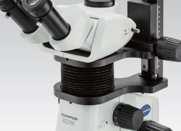
Adjustable Depth of Field with the Built-In AS Zoom Body
Closing the aperture increases the depth of field.
A Wide Array of Accessories Enhance the System for Various Observation and Documentation Methods
The SZX10 microscope’s accessories achieve high performance during image capture and monitor observation. This versatile system can be used for a variety of applications.
Extendable Eyepoint Adjuster
(SZX2-EEPA)
This unit enables users to continuously adjust the height of the eyepoint between 30 mm to 150 mm depending on the user’s eyepoint.
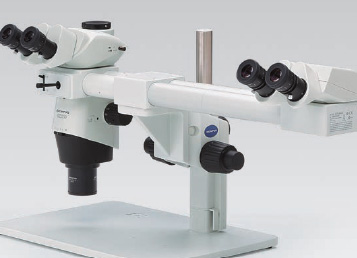
Side-by-Side Discussion Tube
(SZX-SDO2)
Ample distance (650 mm) between the main and secondary observer for easy imaging without disturbing the microscope operation. The color of the built-in pointer can be selected to contrast the specimen.
These tubes enable variable eye points, helping you conduct observations in a natural posture thanks to the tilting head with an incline angle varying between 5º and 45º.
Coaxial Fluorescence Illumination
Stand (SZX-RFA)
This fluorescence unit enables observation of fluorescent proteins introduced into living cells.