We combined our leading-edge technologies—including X Line objectives and True Color LED illumination—to create a fast slide scanner that delivers microscope-quality images onscreen. The SLIDEVIEW™ DX whole slide imaging system’s advanced technology improves the entire digital pathology workflow to help pathologists make diagnoses efficiently with high-quality images they can rely on.
Supports Efficient Diagnosis
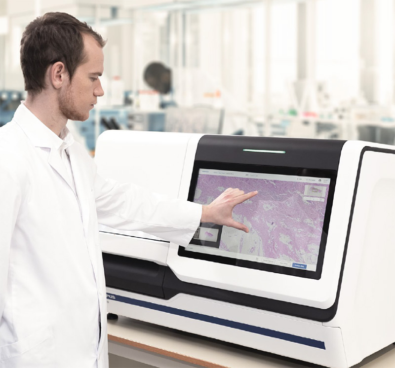
Make your diagnoses with the convenience of digital slide images that you can have confidence in. The SLIDEVIEW DX system uses a dedicated objective lens based on our award-winning X Line™ series to capture images with exceptional flatness and high resolution, enabling seamless stitching and stunning whole slide images that are fully in focus.
Fast, Efficient Pathology Diagnosis Workflow
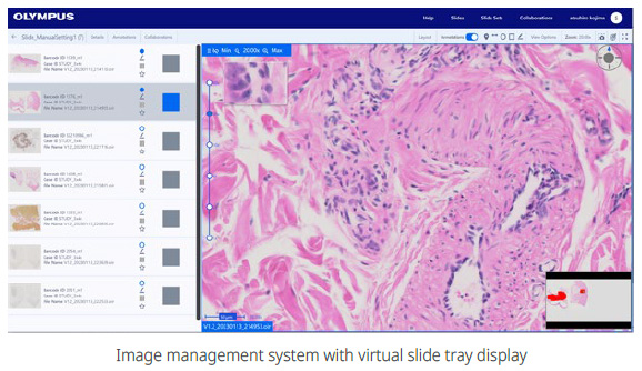
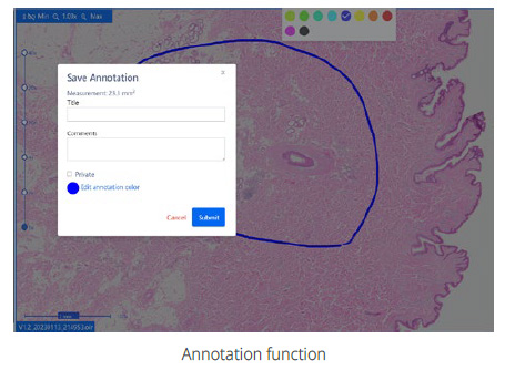
The SLIDEVIEW DX software was developed in collaboration with pathologists, so its layout and functionality will be intuitive and the system easy to learn. The system’s software integrates with your lab information system (LIS) to display the slide and patient information in one convenient window to help you remain focused on making a diagnosis. A virtual slide tray mirrors handling a physical slide on the holder for simple, familiar slide management. View images from the virtual slide tray and easily make measurements and annotations on the digital slide image.
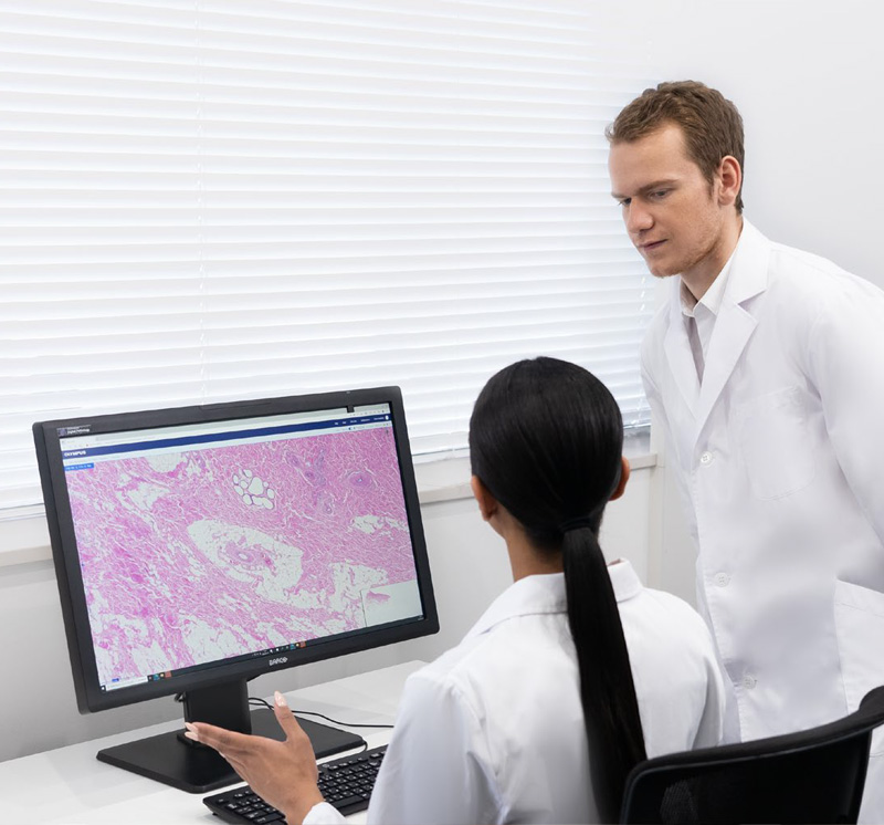
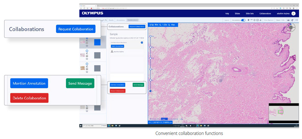
For consultation, the system has convenient collaboration tools that enable you to securely share slide images with other pathologists or experts. Users can access the image management system from a web browser without installing software.
Built for Busy Labs
The SLIDEVIEW DX system makes it easy to scan large numbers of slides. Labs can set up their system based on their preferences. You can configure the system for greater speed and slide quality, giving you flexibility over the scan plan and making it easy to find the right balance of speed and quality. If you need more capacity to run slides, additional scanners can be linked together. For example, using two scanners would allow you to process 300 slides at a time.
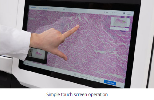
Easy to Operate
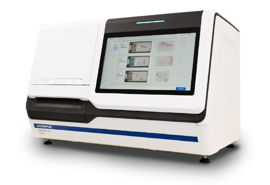
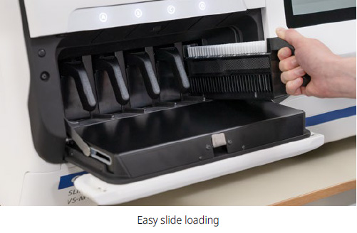
The scanner’s controls are easily accessible from its large touch screen, which is designed to be used while standing. This makes it easy for you to configure the scanner’s controls and walk away. Easily load the slides into the rack and then place them into the scanner. The slides are loaded vertically rather than horizontally to reduce the chance of dropping them. The system walks you through the steps to start scanning. When the scan is complete, you can check the quality of the digital slides using the same touch screen and deliver them directly to the pathologist at the push of a button.
High-Speed Scanning
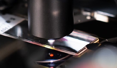
The scanner’s fast, more than 80 slides per hour scan speed enables fast digitization for faster delivery to the pathologist. During the scan, the system’s real-time autofocus enables you to skip the time-consuming focus mapping phase for faster scans.
Reliable
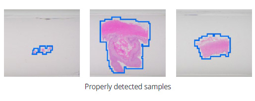
The system’s advanced AI* automatically identifies the tissue on the slide so that only the tissue is scanned. This speeds up the scanning process and minimizes the need for rescans caused by undetected tissue. Easy slide loading Simple touch screen operation Properly detected samples. * Machine learning-based software with regulatory framework.